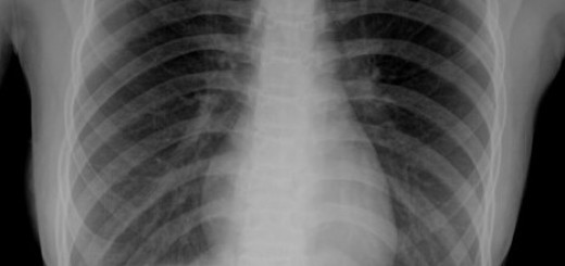
14 year old male presenting with chronic cough for 3 months.


Chest radiograph – Left retrocardiac collapse/consolidation effacing the left hemidiaphragm. The rest of the lung is clear.
CT thorax – confirms a thick rim enhancing fluid collection in the left lower lobe containing pockets of gas and scattered hyperdensities within it. No definite systemic arterial supply is seen leading into the lesion.
abscess, infected congenital malformation such as a congenital pulmonary airway malformation (CPAM), sequestration, bronchogenic cyst, hydatid cyst
A congenital pulmonary airway malformation (CPAM) is a hamartomatous abnormality of the lower respiratory tract, resulting from overgrowth of the terminal bronchioles. Formally known as congenital cystic adenomatoid malformations (CCAM), CPAM is part of a spectrum of bronchopulmonary foregut malformations and account for a quarter of congenital lung abnormalities. It often involves a single lobe, although multi-lobe involvement have been reported.
5 histopathological types reflect size of cysts
- Type 0 (rare): Acinar dysgenesis (incompatible with life)
- Type 1 (65%): 1 or more large (1-10 cm) cysts
- Type 2 (20%): Numerous small (0.5-1.5 cm) cysts
- Type 3 (< 10%): Microcysts (appear solid)
- Type 4 (10%): Large cysts with mass effect (Type 4 indistinguishable from type 1 by imaging)
The size of the CPAM cyst is important in determining the respiratory development/ outcome during the antenatal and perinatal period. Fetuses with large CPAM masses are at risk of developing hydrops and pulmonary hypoplasia. Although there is no satisfactory non-invasive method of determining lung maturity, MRI has shown potential in this regards. On MRI, lung T2W intensity should always be higher than liver, but lower than amniotic fluid. Normal lung intensitiy changes from low to high at around the 26 weeks mark. Lungs that are small and low intensity with ill-defined vessels and interstitium after 26 weeks suggest pulmonary hypoplasia.
- Rosado-de-christenson ML, Stocker JT. Congenital cystic adenomatoid malformation. Radiographics. 1991;11 (5): 865-86
- Rempen A, Feige A, Wunsch P. Prenatal diagnosis of bilateral cystic adenomatoid malformation ofthe lung. JCU 1987; 15:3-8.
- Kuwashima, Shigeko, et al. “Low-intensity fetal lungs on MRI may suggest the diagnosis of pulmonary hypoplasia.” Pediatric radiology 31.9 (2001): 669-672.

Recent Comments